embryo transfer
Grading vs PGT in FET decisions what mattered more?

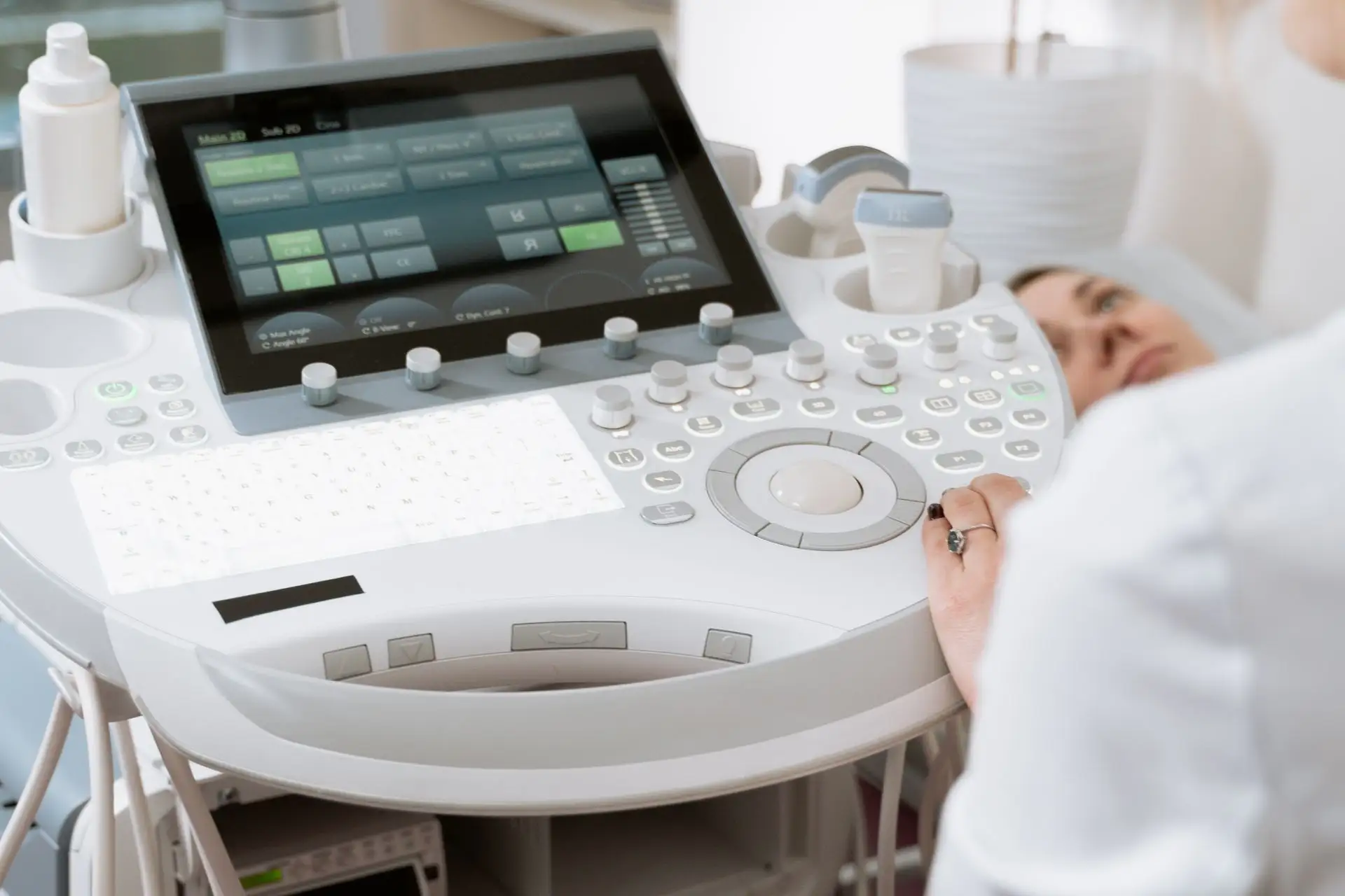
A thin uterine lining, or thin endometrium, is measured via ultrasound as less than 7mm thick around the time of ovulation or embryo transfer. It can make embryo implantation more difficult, potentially contributing to light periods, implantation failure, or early miscarriage. The most common causes are low estrogen, uterine scarring (Asherman’s syndrome), and reduced blood flow to the uterus. In most cases, thin endometrium is treatable with the right protocol.
If a cancelled FET cycle brought you here, you’re dealing with one of the more frustrating fertility obstacles, but effective treatment options exist for most causes.
Your endometrial lining needs to reach an optimal thickness of at least 7–8mm for successful embryo implantation before your frozen embryo transfer. When your lining measures less than 7mm, it’s classified as thin endometrium, a condition that requires targeted intervention before a transfer cycle can proceed. Estimates of its prevalence vary across study populations, but its consistent association with reduced live birth rates in both fresh and frozen transfer cycles makes it a clinically meaningful obstacle to address.
The endometrium is more than just thickness. Your lining needs the right structure, blood flow, and hormonal environment to receive an embryo. Understanding this distinction helps explain why some treatments work better than others, and why the approach must be tailored to the specific cause of thinning.
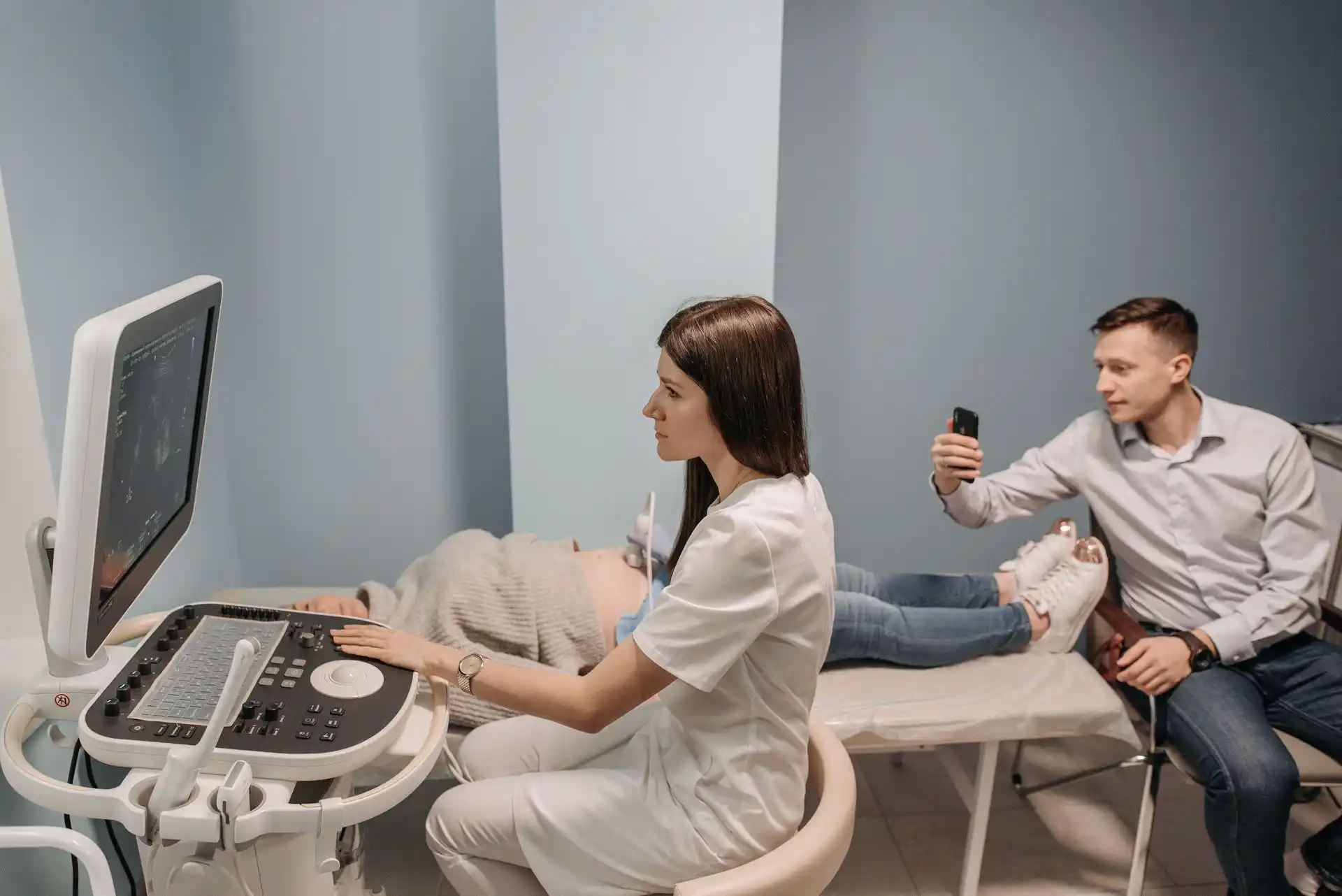
Thin uterine lining often produces no obvious symptoms until fertility testing reveals it. The most common signs include unusually light or short menstrual periods (hypomenorrhea), an absent or thin endometrial stripe on transvaginal ultrasound, repeated implantation failure despite good-quality embryos, and a history of uterine procedures such as D&C, hysteroscopy, or repeated curettage.
Light or absent periods reflect the relationship between estrogen and endometrial growth: low estrogen means less tissue development and less monthly shedding. A thin or absent endometrial stripe on ultrasound (typically below 7mm) is the definitive diagnostic marker and is identified during routine cycle monitoring. Repeated implantation failure despite transferring morphologically normal embryos often points to a receptivity problem rather than an embryo quality issue. Women with a history of uterine infections (PID), intrauterine adhesions (Asherman’s syndrome), or prior use of clomiphene citrate are at elevated risk.
Clomiphene’s anti-estrogenic effect on endometrial tissue is a particularly common cause in PCOS patients who have undergone multiple ovulation induction cycles.
Not sure what’s causing your thin uterine lining? Start with a complete fertility diagnostic at Avida
Several distinct mechanisms can cause inadequate endometrial thickness, and determining which applies to your case is essential before selecting a treatment approach. A comprehensive fertility diagnostic evaluation helps identify the root cause and guides protocol selection.
Identifying the underlying cause directs the treatment. Multiple factors often contribute, requiring a layered protocol that addresses hormonal, vascular, and structural elements simultaneously.
The primary treatments for thin uterine lining are estrogen supplementation (oral, transdermal, or vaginal), progesterone supplementation (timed precisely to the transfer window once target thickness is confirmed), vasodilators (low-dose aspirin 81mg daily; vaginal sildenafil; pentoxifylline), and uterine procedures for structural causes such as hysteroscopy for Asherman’s syndrome. For resistant cases, G-CSF intrauterine infusion and platelet-rich plasma (PRP) therapy have shown clinical benefit in published studies.
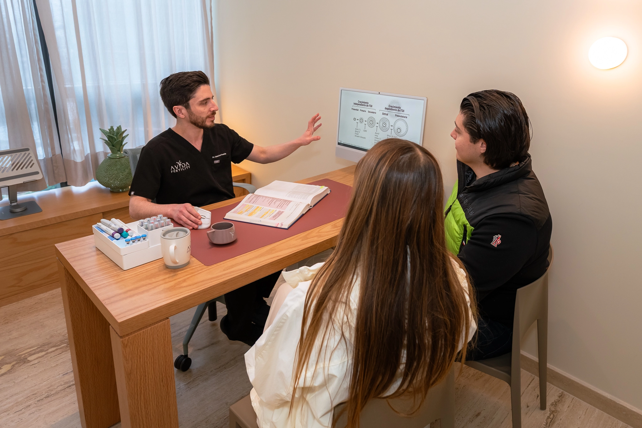
Your body is one of a kind, and so is your treatment. Rely on a highly specialized medical team that explains your options with full transparency, backing every decision with precision technology and genuine, human support from day one.
Estrogen remains the cornerstone treatment for thin endometrium. Different delivery routes offer distinct clinical profiles, and vaginal administration is often the most effective option in resistant cases.
Vaginal administration is particularly effective because estrogen is absorbed directly by uterine tissue, bypassing liver metabolism and achieving higher local uterine concentrations than oral or transdermal routes. For patients who don’t respond to oral or patch protocols, switching to vaginal estradiol is typically the first escalation step.
Progesterone supplementation is introduced once the target thickness and trilaminar pattern are confirmed on ultrasound, completing the transition from estrogen-dominant lining development to the progesterone-supported receptive phase required for embryo implantation. The timing of progesterone start is precisely coordinated with your embryo transfer date and the cryopreservation method used for your embryos. Most FET protocols require 5 to 7 days of progesterone prior to transfer, with dosing continuing through early pregnancy if implantation is confirmed. Progesterone is delivered as vaginal suppositories, intramuscular injections, or oral and transdermal formulations, depending on your clinic’s protocol and your individual tolerance.
G-CSF has emerged as a promising treatment for refractory thin endometrium. This medication, originally developed in oncology, stimulates cell growth and improves blood flow to the uterine lining when infused directly into the uterine cavity.
Published clinical studies on intrauterine G-CSF infusion report increases in endometrial thickness of 2 to 4mm in approximately 60 to 70% of patients who have not responded to standard estrogen therapy. The procedure is performed in an office setting and involves infusing G-CSF directly into the uterine cavity using a thin catheter, without sedation.
For patients where inadequate uterine blood flow is the underlying cause, vasodilator medications are used alongside estrogen to improve uterine perfusion.
Vaginal sildenafil (25mg, 3–4 times daily): inhibits PDE5, promoting nitric oxide-mediated vascular smooth muscle relaxation and increasing uterine arterial blood flow. Administered vaginally for a more direct uterine effect than oral administration.
Pentoxifylline (400mg oral, 2–3 times daily): improves blood rheology by reducing red blood cell aggregation, enhancing microcirculation in uterine tissue and supporting endometrial perfusion.
Vaginal sildenafil and pentoxifylline are used off-label in reproductive medicine under specialist supervision. Their use requires individual clinical evaluation and medical prescription.
Low-dose aspirin (81mg daily) is frequently added to protocols for its antiplatelet effects on uterine microcirculation and is generally well tolerated.
%20Therapy.webp)
PRP therapy uses concentrated growth factors derived from the patient’s own blood platelets to stimulate endometrial regeneration. This approach is particularly relevant for patients who have not responded to standard hormonal therapy and have confirmed thin endometrium despite adequate estrogen exposure.
A study from the UCSF Center for Reproductive Health (Huddleston et al.) involving 46 patients whose prior FET cycles had been cancelled due to endometrial thickness below 6mm demonstrated significant increases in endometrial thickness following intrauterine PRP therapy, with no adverse events reported. Published in the Journal of Assisted Reproduction and Genetics as the first US clinical experience study of this approach, the research showed corresponding improvements in clinical pregnancy outcomes, establishing intrauterine PRP as a viable option for patients with confirmed thin endometrium refractory to standard hormonal therapy.
G-CSF and PRP protocols for thin endometrium are available at Avida at 40–60% of comparable US fertility clinic costs. In the US, uterine PRP is typically offered at $1,500 or more per session as a standalone add-on; at Avida, it is part of an individualized FET preparation protocol. US patients with prior cancelled FET cycles frequently pursue these protocols in Mexico as a cost-effective alternative to what is available domestically. Schedule a free virtual consultation
While medical treatments form the foundation of thin lining therapy, complementary approaches can meaningfully support their effectiveness when combined systematically.
Research indicates that acupuncture can improve FET outcomes by increasing blood flow to the uterus. Studies show that patients receiving acupuncture alongside medical treatment achieve better endometrial thickness compared to medication alone in some protocols. Acupuncture should be coordinated with your fertility team and scheduled around your monitoring cycle.
Specific nutrients support endometrial development and uterine blood flow:
Pomegranate juice is frequently cited in fertility communities for uterine lining support and has a plausible vascular mechanism. Clinical trial evidence is limited, but the risk profile is minimal, making it a reasonable addition during the lining-development phase.

Uterine blood flow is a modifiable factor that responds to lifestyle inputs alongside medical treatment.
Low-dose aspirin (81mg daily) is frequently included in endometrial preparation protocols for its antiplatelet effect on uterine microcirculation. Moderate aerobic exercise (30 minutes, 3–4 times per week) supports systemic circulation without overtaxing the body during a treatment cycle. Heat application (warm abdominal packs or warm baths) promotes local vasodilation and is used in some clinical protocols. Avoiding nicotine is essential: smoking causes vasoconstriction and directly impairs uterine blood flow in a dose-dependent way. Adequate hydration supports blood viscosity and general microcirculation.
These measures complement, but do not replace, medical interventions for significant endometrial thinning. Discuss with your care team which are appropriate alongside your current protocol.
For patients who don’t respond to conventional treatments, advanced options are under active clinical investigation.
While stem cell therapy for thin endometrium remains experimental and is not yet standard clinical practice, early-phase studies have reported promising results in previously treatment-resistant cases. This approach uses mesenchymal stem cells to regenerate endometrial tissue. It is under active clinical investigation and should be discussed with a specialist who actively follows current reproductive medicine research, not approached as an established protocol.
Many fertility clinics now use layered approaches, integrating multiple treatments for maximum effectiveness:
Knowing when to modify your treatment approach is crucial. Consider discussing changes with your fertility team if your lining hasn’t improved after 2–3 cycles of standard estrogen therapy, or if you’ve experienced multiple failed transfers despite achieving adequate thickness. Sometimes the issue is not just numerical thickness but endometrial pattern, receptivity, and hormonal timing.
Clinical experience consistently shows that persistence and protocol adjustment are key to managing refractory thin endometrium. What doesn’t produce results in one cycle may succeed with adjustments in timing, dosage, or combination approach. Most specialists recommend evaluating response after 2–3 cycles before considering more advanced options such as G-CSF, PRP, or, when appropriate, moving toward IVF treatment in Mexico with a comprehensively revised protocol.
If vasodilator therapy has not been tried, that is often the next logical step before transitioning to advanced cellular therapies.
For patients with repeated implantation failure despite adequate endometrial thickness, particularly when structural and hormonal factors have been addressed, Endometrial Receptivity Analysis (ERA) testing can identify the precise implantation window. ERA shifts the question from “Is the lining thick enough?” to “Is the lining receptive at this moment?” and is relevant when cycles are cancelled for thickness reasons repeatedly, even after successful preparation.
Understanding realistic outcomes helps you plan effectively and communicate with your care team. Response to thin endometrium treatment varies meaningfully by underlying cause and patient profile.
Women under 35 with hormonal causes (low estrogen, clomiphene effect) typically see the fastest improvement and highest treatment response rates, often within 1–2 cycles of protocol adjustment. Patients with structural damage (Asherman’s syndrome) or age-related hormonal decline may require more cycles, combination protocols, or additional procedures such as hysteroscopic adhesiolysis before endometrial preparation can proceed effectively.
A 2018 analysis of more than 40,000 embryo transfers (Liu et al., Human Reproduction) found that thin endometrium was associated with lower live birth rates in both fresh and frozen transfer cycles, reinforcing that achieving adequate thickness before FET is clinically meaningful.
Research from Yale School of Medicine found that a thin endometrium may not always prevent IVF success. Some patients achieved live births even with endometrial thickness below the standard 7mm threshold when other parameters were optimal. This reinforces that endometrial adequacy is multifactorial: pattern, receptivity, and embryo quality all matter alongside numerical thickness.
At Avida Fertility in Mexico City, our reproductive specialists, including Dr. Daniel Pascal Pontón (SEP Cédula 13928465), design individualized endometrial preparation protocols for each FET cycle, combining medical treatments (estrogen, G-CSF, PRP, vasodilators) with close Doppler and transvaginal ultrasound monitoring.
We work with patients across all family structures, including single parents by choice, same-sex couples, LGBTQ+ patients using donor gametes, and international patients from the US and Canada, with the same individualized protocol approach.
For US patients, access to G-CSF and PRP protocols is a key differentiator. These advanced therapies are not universally offered at US fertility clinics, and they are available at Avida as part of FET preparation protocols at 40–60% of comparable US fertility clinic costs. Avida Fertility holds COFEPRIS licensing (Mexico’s equivalent of FDA regulation for healthcare facilities) and operates with English-speaking specialists and an internationally equipped embryology lab.
A typical G-CSF or PRP endometrial preparation protocol at Avida runs 2 to 4 weeks of monitoring, timed to coincide with your natural or stimulated cycle. US patients often structure a single trip aligned with monitoring visits and the embryo transfer, or complete early monitoring at a local clinic and travel to Mexico City only for the infusion procedure and transfer itself. Explore the full range of fertility treatments in Mexico available at Avida, or begin with a free virtual consultation to review your previous cycle records before traveling.
Many of our international patients schedule a free virtual consultation before traveling. This allows our team to review previous cycle records, identify protocol gaps, and design a targeted endometrial preparation plan before arrival in Mexico City.
Successful treatment requires careful monitoring and willingness to adjust protocols. Your fertility team will track several parameters throughout your preparation cycle.
Regular transvaginal ultrasounds measure not just thickness but also the pattern and echogenicity of your lining. Blood tests monitor hormone levels to ensure optimal estrogen and progesterone balance. Some clinics also use Doppler ultrasound to assess blood flow to the uterine arteries.
Communicate with your medical team about any concerns or side effects. Treatment adjustments are common and often necessary for achieving the best outcomes. The willingness to adapt distinguishes effective thin endometrium management from repeated unsuccessful cycles.
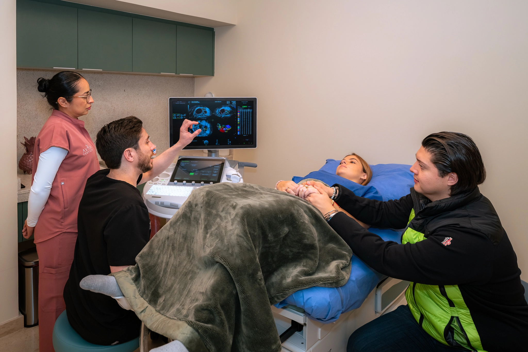
We know every story is different and that taking the first step takes courage. Our medical team takes the time to assess your overall health and walk you through your options step by step, in a safe space and with complete transparency.
A trilaminar endometrial pattern, visible on transvaginal ultrasound as three distinct layers, is associated with better implantation outcomes than a homogeneous (uniform) pattern of equal thickness. Most fertility clinics prefer to see a trilaminar pattern alongside a thickness of at least 7mm before proceeding with embryo transfer. A homogeneous or non-trilaminar lining may indicate inadequate estrogen stimulation or reduced uterine receptivity, even if the numerical thickness appears sufficient.
On ultrasound, the trilaminar pattern appears as an outer hypoechoic (dark) layer, a central echogenic (bright) line, and an inner hypoechoic layer. This three-layer structure reflects adequate estrogenic stimulation and proper structural organization of the endometrium. Some studies have found that patients with a 7mm homogeneous lining may have lower implantation rates than patients with a 6.5mm trilaminar lining, reinforcing that pattern quality is not secondary to thickness.
The same estrogen protocols used to increase endometrial thickness also promote the development of a trilaminar pattern. If your ultrasound report describes your lining as “homogeneous” rather than trilaminar at adequate thickness, this is worth discussing with your specialist as it may indicate suboptimal estrogenic stimulation despite numerical adequacy.
Struggling with thin endometrium? Our specialists have solutions. Contact Avida Fertility.

Once your lining reaches optimal thickness and pattern, timing becomes the critical next variable. The transition from estrogen-dominant to progesterone-supported environment must be precisely coordinated with your embryo transfer.
Your medical team will provide specific instructions about medication timing, activity restrictions, and what to expect during the transfer process. Following these guidelines carefully maximizes your chances of a successful transfer.
A trilaminar endometrial pattern, visible on transvaginal ultrasound as three distinct layers, is associated with better implantation outcomes than a homogeneous (uniform) pattern of equal thickness. Most fertility clinics prefer to see a trilaminar pattern alongside a thickness of at least 7mm before proceeding with embryo transfer. A homogeneous or non-trilaminar lining may indicate inadequate estrogen stimulation or reduced uterine receptivity, even if the numerical thickness appears sufficient.
On ultrasound, the trilaminar pattern appears as an outer hypoechoic (dark) layer, a central echogenic (bright) line, and an inner hypoechoic layer. This three-layer structure reflects adequate estrogenic stimulation and proper structural organization of the endometrium. Some studies have found that patients with a 7mm homogeneous lining may have lower implantation rates than patients with a 6.5mm trilaminar lining, reinforcing that pattern quality is not secondary to thickness.
The same estrogen protocols used to increase endometrial thickness also promote the development of a trilaminar pattern. If your ultrasound report describes your lining as “homogeneous” rather than trilaminar at adequate thickness, this is worth discussing with your specialist as it may indicate suboptimal estrogenic stimulation despite numerical adequacy.
Struggling with thin endometrium? Our specialists have solutions.


In a natural cycle, the endometrium grows approximately 1mm per day during days 6–14, reaching 7–14mm by ovulation. With estrogen therapy, most patients achieve a target thickness of 7–10mm within 10–21 days, though some require longer protocols or dose adjustments. The “48-hour” thickening commonly searched online is not a realistic clinical expectation; gradual daily growth is the normal pattern. After a single insufficient cycle, most specialists recommend 2–3 full cycles before concluding that a protocol isn’t working.
The ability to permanently improve thin lining depends on the underlying cause. Hormonal causes often respond well to treatment and may not require ongoing intervention once addressed. Structural damage such as Asherman’s syndrome may require ongoing hormonal support during each cycle. Many patients maintain good lining thickness with appropriate hormone protocols throughout their treatment plan.
Most fertility clinics aim for at least 7–8mm with a trilaminar pattern on ultrasound. Quality and pattern matter as much as numerical thickness. Some patients achieve successful transfers at 6.5mm with an optimal trilaminar pattern, and research from Yale School of Medicine suggests that thin endometrium may not always prevent success when other parameters are favorable.
Standard estrogen therapy has minimal risks when properly monitored. Newer treatments like G-CSF and PRP have shown good safety profiles in clinical studies, though long-term data is still being collected. Off-label vasodilators (sildenafil, pentoxifylline) require specialist supervision and individual clinical evaluation. Combination protocols involving multiple agents should always be managed by an experienced reproductive specialist.
This decision should be made with your fertility team based on your thickness measurement, endometrial pattern, and embryo quality. Research from Yale School of Medicine suggests that thin endometrium may not always hinder IVF success; some patients achieve live births below standard thresholds when other parameters are optimal. There are also cases where cancellation and protocol revision is the more appropriate clinical choice.
Yes. Chronic stress can affect hormone production and uterine blood flow, potentially disrupting endometrial development. Cortisol excess from chronic stress interferes with estrogen signaling. Stress management techniques, including mindfulness, moderate exercise, and improving sleep quality, can be a beneficial addition to your treatment plan.
.png)
.svg)


.svg)
.svg)